Top 5 Myths About MRI Scans

Top 5 Myths About MRI Scans: Debunking Common Misconceptions

Introduction

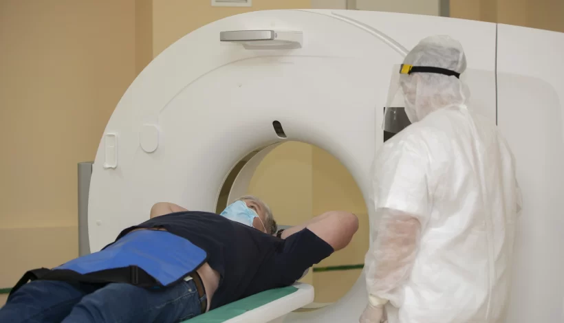
Magnetic Resonance Imaging (MRI) is one of the most advanced diagnostic imaging tools used in modern medicine. Despite its accuracy and safety, many patients still feel anxious about undergoing an MRI scan due to widespread myths and misconceptions. These myths often prevent people from seeking timely medical help, leading to unnecessary delays in diagnosis and treatment.

In this blog, we will debunk the Top 5 Myths About MRI Scans with clear explanations to help patients feel confident and informed before their procedure.

What is an MRI Scan?

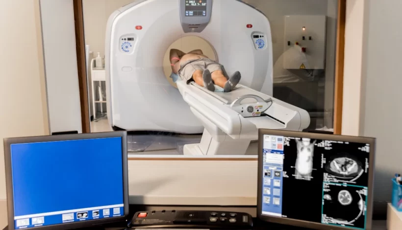
An MRI scan uses a powerful magnetic field, radio waves, and a computer to create detailed images of the inside of the body. Unlike X-rays and CT scans, MRI does not use ionizing radiation, making it a safe and effective tool for diagnosing a wide range of conditions.

MRI is commonly used for:

  • Brain and spinal cord imaging
  • Joint, ligament, and soft tissue evaluation
  • Detecting tumors and cancers
  • Heart and blood vessel imaging
  • Abdominal and pelvic organ scans

Now, let’s break down the Top 5 Myths About MRI Scans and separate fact from fiction.

Myth 1: MRI Scans Are Unsafe Because of Radiation

One of the most common misconceptions is that MRI scans expose patients to harmful radiation, similar to X-rays or CT scans.

The Truth:

  • MRI does not use radiation at all. Instead, it relies on magnetic fields and radiofrequency waves.
  • The magnetic field temporarily realigns hydrogen atoms in the body. When these atoms return to their normal position, they emit signals that are captured to create images.
  • Unlike CT scans, which use a small dose of ionizing radiation, MRI is completely free of radiation exposure.

Why This Matters:

This makes MRI scans particularly safe for patients of all ages, including children and pregnant women (with certain precautions).

Myth 2: MRI Scans Are Only for Serious Conditions

Some patients believe that an MRI scan is only prescribed for severe illnesses like cancer or brain tumors.

The Truth:

  • MRI scans are versatile and can be used for both serious and routine medical evaluations.
  • Doctors often recommend MRI for:
    • Back pain and slipped disc
    • Sports injuries (ligaments, tendons, cartilage)
    • Chronic headaches or migraines
    • Knee or shoulder injuries
    • Abdominal pain and pelvic conditions

Why This Matters:

Even if you are not dealing with a life-threatening condition, your doctor may order an MRI scan to get a clearer view of soft tissues, which cannot be seen as well with X-rays or ultrasounds.

Myth 3: MRI Scans Are Painful and Dangerous

Many patients avoid MRI scans because they fear it might hurt or cause side effects.

The Truth:

  • An MRI scan is completely painless. You won’t feel the magnetic field or radio waves.
  • The only discomfort comes from:
    • Staying still during the scan (usually 15–60 minutes)
    • Loud noises from the machine (earplugs or headphones are provided)
  • MRI is non-invasive and does not cause long-term side effects.

Safety Considerations:

  • Patients with certain implants (such as pacemakers, cochlear implants, or metal fragments) must inform their doctor beforehand, as the magnetic field may interfere.
  • Otherwise, MRI is one of the safest diagnostic imaging procedures available.

Myth 4: People with Claustrophobia Cannot Have an MRI

Claustrophobia, or fear of enclosed spaces, is a common concern when it comes to MRI scans. Some patients believe they cannot undergo MRI if they have anxiety in confined spaces.

The Truth:

  • While MRI scanners are enclosed tubes, there are solutions for patients with claustrophobia:
    • Open MRI machines: Wider and less enclosed, designed for comfort.
    • Sedation options: Mild sedatives may be used to help patients relax.
    • Music or communication systems: Many centers provide headphones with music or allow constant communication with the technologist.

Why This Matters:

Modern MRI technology is patient-friendly. If you are claustrophobic, you can still undergo MRI safely with the right support.

Myth 5: MRI Scans Take Too Long and Are Inconvenient

Some people think an MRI scan requires hours of preparation and scanning, making it too inconvenient for their schedule.

The Truth:

  • A typical MRI scan takes 15 to 60 minutes, depending on the body part being imaged.
  • Preparation is minimal:
    • Remove metallic objects (jewelry, watches, belts).
    • Change into a hospital gown if required.
    • Answer safety questions about implants or medical devices.
  • Patients can usually return to normal activities immediately after the scan.

Why This Matters:

MRI scans are quick, efficient, and non-invasive. They provide valuable diagnostic information that can save time in long-term treatment planning.

Summary Table: Myths vs. Facts About MRI Scans

MythFact
MRI uses harmful radiationMRI uses magnetic fields, not radiation
MRI is only for serious diseasesMRI is used for both routine and critical conditions
MRI is painful and riskyMRI is safe, painless, and non-invasive
Claustrophobic patients cannot undergo MRIOptions like open MRI and sedation are available
MRI takes too long and is inconvenientMost MRI scans take only 15–60 minutes

Benefits of MRI Scans Over Other Imaging Techniques

To better understand why MRI is often recommended, here’s a quick comparison:

Imaging MethodRadiation ExposureBest ForLimitations
X-rayYesBones, fracturesLimited soft tissue detail
CT ScanYes (low dose)Trauma, chest/abdominal imagingRadiation exposure
UltrasoundNoPregnancy, abdominal organsLimited in deep tissues
MRINoBrain, spine, joints, soft tissues, tumorsLonger scan time, not suitable for patients with certain implants

Tips for Patients Before an MRI Scan

To make your MRI experience smoother:

  • Inform your doctor if you have implants, pacemakers, or metal fragments.
  • Avoid wearing metallic objects (jewelry, zippers, belts).
  • Stay relaxed – MRI scans are safe and painless.
  • Ask about sedation or open MRI if you have claustrophobia.
  • Follow any fasting instructions if contrast dye is required.

Why Choose Ace Imaging Centre for MRI Scans?

At Ace Imaging Centre, we understand that patients may feel anxious about MRI scans due to common myths and misconceptions. Our facility is designed to ensure:

  • State-of-the-art MRI technology for accurate results
  • Patient comfort with open MRI and relaxation options
  • Experienced radiologists and technicians who guide you at every step
  • Quick reporting and diagnosis to help your doctor plan treatment faster

When you choose Ace Imaging Centre, you are choosing safety, accuracy, and care.

Final Thoughts

MRI scans are one of the safest and most reliable diagnostic tools in modern medicine. Unfortunately, myths about MRI scans often create unnecessary fear among patients. By understanding the facts, you can approach your MRI appointment with confidence.

Remember:

  • MRI does not use radiation.
  • It is safe, painless, and effective.
  • Claustrophobia and convenience concerns can be easily managed.

At Ace Imaging Centre, we are committed to making your MRI experience stress-free and accurate.

    Book an appointment

    Fill form and get discount. Check Offers

    DOTA Scan vs PET Scan

    DOTA Scan vs PET Scan – Complete Guide for Patients

    Introduction

    Medical imaging has revolutionised the way doctors diagnose and treat diseases. Two commonly discussed scans are the DOTA scan and the PET scan. While both involve advanced nuclear medicine techniques, they are not the same. Understanding the difference between a DOTA scan vs PET scan can help patients feel informed and confident before undergoing either procedure.

    This guide explains in detail what each scan is, how they work, when they are recommended, and their benefits, so you can make informed healthcare decisions with your doctor.

    What Is a PET Scan?

    PET scan stands for Positron Emission Tomography scan. It is an advanced imaging test that uses a small amount of radioactive tracer to detect abnormalities in the body at the cellular level.

    • Purpose: Detect metabolic changes in tissues before structural changes occur.
    • Common Uses:
      • Cancer detection, staging, and treatment monitoring
      • Heart disease evaluation
      • Brain disorders (e.g., Alzheimer’s, epilepsy)
    • Tracer Used: Commonly FDG (Fluorodeoxyglucose)

    How It Works:

    1. A radioactive tracer is injected into the bloodstream.
    2. The tracer travels to target organs or cells.
    3. The PET scanner detects gamma rays emitted from the tracer, creating detailed images of body function.

    What Is a DOTA Scan?

    A DOTA scan is a specialised type of PET scan used primarily to detect neuroendocrine tumours (NETs).

    • Full Form: DOTA refers to DOTATATE or DOTATOC, which are molecules that bind to somatostatin receptors on certain tumour cells.
    • Purpose: Specifically targets tumours with somatostatin receptors.
    • Tracer Used: Gallium-68 DOTATATE or Gallium-68 DOTATOC.

    How It Works:

    1. A radiolabelled DOTA compound is injected into the patient.
    2. It binds to somatostatin receptors present on certain tumours.
    3. PET/CT scanning captures high-contrast images showing tumour locations and spread.

    DOTA Scan vs PET Scan – Key Differences

    While both scans use PET technology, there are important differences in the tracer used, the target, and the clinical applications.

    FeaturePET Scan (FDG)DOTA Scan (Ga-68 DOTATATE)
    Main PurposeDetects abnormal metabolism in various tissuesDetects neuroendocrine tumours with somatostatin receptors
    Tracer UsedFDG (fluorodeoxyglucose)Ga-68 DOTATATE or DOTATOC
    Best ForCancer, brain, heart diseasesNeuroendocrine tumours (NETs)
    SpecificityGeneral metabolic imagingHighly specific to NETs
    PreparationFasting 4–6 hours beforeSimilar fasting; sometimes additional instructions for NET patients
    Scan Time20–45 minutes20–45 minutes

    When Doctors Recommend a PET Scan

    A PET scan may be recommended for:

    • Detecting cancer early
    • Determining the extent (stage) of cancer
    • Checking if treatment is working
    • Evaluating brain disorders like Alzheimer’s
    • Assessing heart muscle damage after a heart attack

    When Doctors Recommend a DOTA Scan

    A DOTA scan is usually ordered for:

    • Diagnosing suspected neuroendocrine tumours
    • Determining tumour spread (metastasis)
    • Evaluating treatment response in NETs
    • Locating small or hidden NETs not seen on other scans

    Procedure – What Patients Can Expect

    PET Scan Procedure

    1. Fasting for 4–6 hours.
    2. Injection of FDG tracer.
    3. Rest period to allow tracer distribution.
    4. Lying still inside the PET/CT scanner while images are taken.

    DOTA Scan Procedure

    1. Similar fasting guidelines.
    2. Injection of Ga-68 DOTATATE tracer.
    3. Waiting period (usually 45–60 minutes) for tracer uptake.
    4. PET/CT scanning.

    Advantages of PET Scan

    • Detects disease at an early stage.
    • Can assess how well treatment is working.
    • Useful in multiple medical fields (oncology, neurology, cardiology).

    Advantages of DOTA Scan

    • High accuracy for detecting NETs.
    • Can find tumours too small for conventional imaging.
    • Guides targeted therapy decisions.

    Limitations & Risks

    PET Scan Limitations:

    • False positives if inflammation is present.
    • Limited use for tumours with low glucose metabolism.

    DOTA Scan Limitations:

    • Only useful for tumours with somatostatin receptors.
    • Not for all cancer types.

    Risks for Both:

    • Small exposure to radiation.
    • Rare allergic reactions to tracer.
    • Mild discomfort at injection site.

    Preparation Tips for Patients

    • Follow fasting instructions carefully.
    • Avoid strenuous exercise 24 hours before the scan.
    • Inform your doctor about all medications and supplements.
    • For diabetic patients: Special scheduling may be required.

    DOTA Scan vs PET Scan – Choosing the Right Test

    The decision depends on:

    • Type of disease suspected
    • Tumour biology
    • Doctor’s assessment based on medical history and prior tests

    Example:
    If a doctor suspects lung cancer, a standard FDG PET scan may be chosen.
    If a patient has symptoms suggestive of a neuroendocrine tumour, a DOTA scan is more accurate.

    Cost & Availability

    • PET Scan: More widely available; cost varies by hospital and region.
    • DOTA Scan: Available in specialised imaging centres like Ace Imaging Centre; cost may be higher due to tracer specificity.

    Why Choose Ace Imaging Centre for PET or DOTA Scans?

    At Ace Imaging Centre, patients benefit from:

    • State-of-the-art PET/CT scanners
    • Experienced nuclear medicine specialists
    • Accurate diagnosis for better treatment planning
    • Comfortable and patient-friendly environment

    Our team ensures every scan is performed with utmost care, accuracy, and safety.

    Frequently Asked Questions

    Q1: Is a DOTA scan the same as a PET scan?
    No. A DOTA scan is a type of PET scan using a specific tracer for neuroendocrine tumours.

    Q2: Which is more accurate – DOTA scan or PET scan?
    Accuracy depends on the disease. For NETs, DOTA scans are superior. For general cancers, FDG PET scans are preferred.

    Q3: Is the radiation exposure dangerous?
    Both involve low radiation doses, generally considered safe under medical supervision.

    Q4: Can I eat before the scan?
    Fasting for 4–6 hours is usually required for both scans.

    Q5: How long do results take?
    Results are typically available within 24–48 hours.

    Conclusion

    While both scans use PET imaging technology, the DOTA scan vs PET scan debate is really about the right tool for the right job. A standard PET scan is versatile and used across many diseases, while a DOTA scan is highly specific for neuroendocrine tumours.

    At Ace Imaging Centre, we ensure patients get the correct scan based on their unique condition, leading to more accurate diagnoses and better treatment outcomes.

      Book an appointment

      Fill form and get discount. Check Offers

      Introduction to Pathology

      Introduction to Pathology: What It Is, How It Works, and Why It Matters

      Introduction to Pathology

      Pathology is the cornerstone of modern medicine. It forms the scientific basis for understanding diseases, diagnosing health conditions, and guiding clinical treatments. At Ace Imaging Center, we believe that every accurate diagnosis begins with high-quality pathology services. Whether it’s analyzing blood, tissue, or body fluids, pathology provides vital insights into the causes and progression of diseases.

      This blog serves as a comprehensive introduction to pathology, exploring its definition, branches, diagnostic methods, and its crucial role in patient care.

      What is Pathology?

      Pathology is the study of disease its causes, development, and effects on the body. It involves the examination of organs, tissues, bodily fluids, and cells to understand the nature of diseases and guide treatment decisions.

      Key Functions of Pathology:

      • Diagnose diseases like infections, cancer, or autoimmune conditions
      • Monitor the progression of chronic illnesses
      • Support preventive healthcare through early detection
      • Guide treatment plans and assess response to therapies

      What is a Pathology Test?

      A pathology test refers to any laboratory analysis performed on bodily samples—such as blood, urine, stool, tissues, or swabs—to detect abnormalities, monitor health, and diagnose diseases.

      Common Purposes of Pathology Tests:

      • Identifying infections (bacterial, viral, fungal)
      • Checking organ function (liver, kidney, thyroid)
      • Detecting cancerous cells
      • Measuring blood sugar or cholesterol levels
      • Monitoring medication effects

      Common Types of Pathology Tests:

      Test TypeExamplesPurpose
      Blood TestsCBC, LFT, KFT, Lipid ProfileGeneral health, organ function
      Urine TestsRoutine Urinalysis, CultureKidney function, infections
      Stool TestsOccult Blood, ParasitesDigestive health, GI bleeding
      BiopsiesBreast, liver, prostate tissue analysisCancer diagnosis
      CytologyPap smear, FNACDetecting cancer or abnormal cells
      MicrobiologySputum, blood, urine culturesIdentify infectious agents
      Molecular TestsPCR, genetic mutation panelsDNA/RNA analysis for precise diagnosis

      At Ace Imaging Center, all tests are performed using modern equipment and interpreted by expert pathologists to ensure accuracy.

      What is a Pathology Lab?

      A pathology lab (also called a diagnostic laboratory) is a specialized medical facility where tests are conducted on clinical specimens to obtain information about a patient’s health.

      Services Offered by a Pathology Lab:

      • Sample collection and preparation
      • Laboratory testing and analysis
      • Result interpretation by pathologists
      • Diagnostic support to clinicians

      Key Staff in a Pathology Lab:

      • Pathologists: Medical doctors who interpret test results
      • Lab Technicians: Perform testing procedures and maintain equipment
      • Phlebotomists: Collect blood and other samples
      • Microbiologists: Analyze infections
      • Molecular Biologists: Conduct advanced DNA-based testing

      At Ace Imaging Center, our NABL-accredited lab ensures safe, hygienic, and efficient testing with digital reporting and expert consultation.

      Branches of Pathology

      Pathology is a diverse field, divided into several branches based on the type of investigation and sample material. Each branch plays a specific role in medical diagnosis and research.

      BranchFocus Area
      Anatomical PathologyExamines tissues and organs for disease (e.g., biopsies, autopsies)
      Clinical PathologyAnalyzes blood, urine, and other body fluids to detect disease
      HematologyStudies blood and blood-forming tissues (e.g., anemia, leukemia)
      HistopathologyMicroscopic analysis of tissue samples
      CytopathologyStudies individual cells (e.g., Pap smear, FNAC)
      MicrobiologyIdentifies infections caused by bacteria, viruses, fungi, and parasites
      ImmunologyInvestigates immune responses and autoimmune diseases
      Molecular PathologyExplores genetic and molecular alterations in cells, especially in cancer diagnosis

      Each of these specialties is crucial in delivering comprehensive and accurate diagnostic services.

      Importance of Pathology in Modern Healthcare

      Pathology plays an indispensable role in diagnosing, managing, and preventing diseases. Without it, clinicians would be operating without a clear understanding of the patient’s condition.

      Why Pathology Matters:

      • Early Diagnosis: Detects diseases in their earliest stages, improving treatment outcomes.
      • Treatment Planning: Helps doctors choose the most effective treatments based on lab reports.
      • Prognosis Estimation: Predicts the likely course and outcome of a disease.
      • Monitoring Progress: Tracks changes in disease progression or response to treatment.
      • Screening: Identifies individuals at risk for specific conditions (e.g., cancer screening).

      Types of Pathology Tests

      There are numerous tests conducted under the field of pathology. These tests are typically categorized based on the sample type and the condition being investigated.

      Common Pathology Test Categories:

      1. Blood Tests
        • CBC (Complete Blood Count)
        • Blood Sugar Levels
        • Liver and Kidney Function Tests
        • Lipid Profile
        • Thyroid Function Tests
      2. Urine Tests
        • Routine Urinalysis
        • Urine Culture
        • Microalbumin Test
      3. Stool Tests
        • Occult Blood
        • Parasite Detection
      4. Biopsy and Histopathology
        • Tissue examination for cancer and other disorders
      5. Cytology
        • Pap smear
        • Fine Needle Aspiration Cytology (FNAC)
      6. Microbiological Culture Tests
        • Blood culture
        • Sputum culture
        • Wound swab culture
      7. Molecular and Genetic Testing
        • PCR tests
        • Cancer gene panels
        • BRCA testing for breast cancer

      Each test is performed with the highest standards of accuracy and reliability at Ace Imaging Center.

      The Pathology Testing Process: From Sample Collection to Report

      At Ace Imaging Center, the pathology process is handled with utmost precision to ensure accurate and timely results. Here’s a step by step breakdown of how it works:

      1. Sample Collection

      • Blood, urine, stool, tissue, or other biological materials are collected by trained phlebotomists or clinicians.
      • Proper labeling and handling are critical to avoid contamination or mix-ups.

      2. Sample Transportation

      • Samples are safely transported to the lab under controlled conditions to preserve their integrity.

      3. Laboratory Analysis

      • Advanced machines and microscopes analyze the samples based on the test ordered.
      • Pathologists, lab technicians, and medical microbiologists interpret findings.

      4. Result Interpretation

      • Qualified pathologists review results to confirm diagnoses.
      • The report includes findings, reference ranges, and comments for physicians.

      5. Report Delivery

      • Final reports are shared with the physician and patient digitally or in print.
      • Turnaround time depends on the complexity of the test.

      Who Performs Pathology Tests?

      Pathology tests are conducted by a team of highly trained healthcare professionals, each playing a vital role in ensuring diagnostic accuracy.

      Key Personnel in a Pathology Lab:

      • Pathologists: Doctors specialized in diagnosing disease through lab data and tissue analysis.
      • Lab Technicians: Skilled professionals who handle sample processing and test execution.
      • Microbiologists: Experts in detecting infections and antibiotic sensitivities.
      • Phlebotomists: Trained staff for collecting blood and other specimens from patients.
      • Molecular Biologists: Specialists in DNA/RNA testing and advanced cancer diagnostics.

      Role of Pathology in Cancer Diagnosis

      One of the most vital applications of pathology is in oncology (cancer diagnosis and treatment). Pathologists examine tissue biopsies to:

      • Confirm the presence of cancer
      • Identify cancer type (e.g., carcinoma, lymphoma)
      • Determine grade and stage
      • Guide oncologists in selecting appropriate therapies
      • Predict prognosis and risk of recurrence

      Example Tests in Cancer Pathology:

      TestPurpose
      Biopsy with HistopathologyConfirms type and aggressiveness of cancer
      Immunohistochemistry (IHC)Identifies specific tumor markers
      Molecular TestingDetects gene mutations (e.g., EGFR, KRAS)
      Liquid BiopsyNon-invasive detection of tumor DNA in blood

      Pathology and Preventive Healthcare

      Preventive medicine relies heavily on routine pathology screenings to detect issues before symptoms arise. This proactive approach helps in lowering disease burden and healthcare costs.

      Key Preventive Tests:

      • Annual health check-ups (CBC, blood sugar, lipids)
      • Cancer screenings (Pap smear, PSA test, mammography)
      • Thyroid and hormone levels
      • Diabetes markers (HbA1c)
      • Kidney function monitoring

      Digital Pathology: The Future of Diagnostics

      With the advent of technology, pathology is rapidly evolving. Digital pathology allows high-resolution images of tissue samples to be analyzed and shared remotely. This enhances:

      • Speed of diagnosis
      • Remote consultations (telepathology)
      • AI-assisted interpretation
      • Archival and tracking of patient data

      Ace Imaging Center is committed to adopting the latest advancements, ensuring world-class care.

      Why Choose Ace Imaging Center for Pathology Services?

      At Ace Imaging Center, we take pride in offering comprehensive pathology services backed by experienced professionals and advanced technologies.

      What Sets Us Apart?

      • NABL-accredited laboratory
      • State-of-the-art testing equipment
      • Rapid report turnaround
      • Expert team of certified pathologists and technicians
      • Hygienic and safe sample collection practices
      • Digital reports for easy access
      • Customizable health packages
      • Affordable rates with transparent billing

      We ensure that every report you receive is accurate, timely, and easy to understand because your health deserves nothing less.

      Frequently Asked Questions (FAQs)

      1. What is the difference between pathology and radiology?

      Pathology involves lab tests on tissues and body fluids, while radiology uses imaging like X-rays, CT scans, and MRIs to view internal body structures.

      2. How long does it take to get pathology test results?

      Basic tests may take a few hours, while complex ones like biopsies can take 2–5 days depending on the analysis needed.

      3. Are pathology tests painful?

      Most pathology tests are non-invasive or involve minor procedures like blood draws or swabs, which cause minimal discomfort.

      4. Can I get my pathology report online?

      Yes, Ace Imaging Center provides secure digital reports accessible through email or our online portal.

      5. Do I need a doctor’s prescription for pathology tests?

      Some tests require a doctor’s referral, but preventive screenings or health packages can often be booked directly.

      Final Thoughts

      This introduction to pathology highlights just how central this field is to every stage of patient care from prevention to diagnosis and treatment. Whether you’re undergoing a routine blood test or a complex biopsy, pathology is the key to unlocking critical insights about your health.

      Choosing a reliable and experienced diagnostic partner like Ace Imaging Center ensures you get the care, attention, and accuracy you deserve. Our mission is to empower patients and physicians with reliable lab support that makes all the difference.

      Book Your Test Today at Ace Imaging Center
      For safe, accurate, and affordable pathology services in Mumbai, trust the experts at Ace Imaging Center. Call us or visit our website to schedule your appointment.

        Book an appointment

        Fill form and get discount. Check Offers

        PET Scan Centres in Mumbai

        2 Years of Diagnostic Excellence

        Introduction:

        We’re thrilled to share a proud milestone — ACE PET-CT has completed 2 successful years of providing world-class diagnostic services in Mumbai and the surrounding regions. Over the past two years, we’ve had the privilege of serving more than 5,000 patients, offering precise and timely PET scan, CT scan, and MRI scan services to aid in early detection and effective treatment planning.

        As one of the region’s most advanced PET scan centres, ACE Imaging Centre is dedicated to accuracy, patient comfort, and medical excellence — a combination that has made us a trusted name among doctors and patients alike.

        The Power of Medical Imaging: PET, CT & MRI Scans Explained

        Medical imaging plays a central role in modern diagnosis and treatment. At ACE Imaging Centre, we offer a full range of advanced imaging services, including:

        PET Scan (Positron Emission Tomography)

        PET scans provide functional imaging by tracking metabolic activity in organs and tissues. It’s particularly effective in:

        • Detecting and staging cancer
        • Monitoring cancer treatment
        • Evaluating brain disorders like epilepsy and Alzheimer’s
        • Assessing heart conditions and blood flow

        CT Scan (Computed Tomography)

        A CT scan uses X-rays and computer processing to create detailed cross-sectional images of bones, blood vessels, and soft tissues. It helps in:

        • Detecting fractures, infections, or tumors
        • Guiding surgical planning
        • Evaluating internal injuries and bleeding

        MRI Scan (Magnetic Resonance Imaging)

        MRI uses powerful magnets and radio waves to create highly detailed images, especially of soft tissues. It is the preferred modality for:

        • Brain and spine imaging
        • Joint and musculoskeletal evaluations
        • Detailed organ imaging (liver, kidneys, uterus, prostate, etc.)

        By offering all three imaging modalities under one roof, we provide comprehensive diagnostic solutions to support every medical specialty — from oncology to neurology, cardiology, orthopaedics, and more.

        Why ACE PET-CT Is a Top Choice Among PET Scan Centres

        Choosing a diagnostic centre isn’t just about availability — it’s about accuracy, safety, speed, and trust. Here’s why ACE Imaging Centre is considered a top performer among PET scan centres, and a reliable destination for MRI and CT scan services as well:

        1. Advanced Imaging Equipment

        • High-resolution digital PET-CT scanners for accurate metabolic and structural imaging
        • 128-slice CT scanners for rapid, detailed cross-sectional images
        • 3T MRI systems for ultra-clear soft tissue scans

        We invest in the latest technology to ensure better clarity, faster scanning, and reduced radiation exposure for patients.

        2. Experienced Medical Team

        • Board-certified radiologists and nuclear medicine specialists with years of experience
        • Trained technologists and support staff ensuring smooth scan procedures
        • Collaborative case discussions for complex diagnoses

        3. Quick Turnaround & Digital Access

        • Same-day or next-day scan reports
        • Digital sharing with doctors and hospitals
        • Secure patient portal for accessing scan images and reports

        4. Comfort-Focused Patient Experience

        • Clear pre-scan instructions and counseling
        • Clean, comfortable waiting and scan rooms
        • Gentle handling and support throughout your visit

        Our focus on compassionate care makes a real difference for patients who may be anxious or undergoing medical treatment.

        Comprehensive Imaging Services Under One Roof

        Here’s a quick breakdown of the diagnostic imaging services available at ACE Imaging Centre:

        ServiceApplications
        Whole Body PET-CTOncology staging, treatment response
        FDG PET-CT ScanCommonly used for cancer detection
        PSMA PET-CTProstate cancer evaluation
        DOTA PET-CTFor neuroendocrine tumors
        Brain PET ScanAlzheimer’s, epilepsy, brain tumors
        CT Chest/AbdomenLung, liver, and abdominal imaging
        CT Coronary AngiographyNon-invasive heart evaluation
        MRI Brain/SpineNeurological disorders, stroke, disc herniation
        MRI JointsSports injuries, arthritis, ligament tears
        Contrast ImagingCT/MRI with contrast for enhanced clarity

        Whether you need a PET scan for cancer detection, a CT scan after trauma, or an MRI scan for chronic pain, we provide accurate imaging that physicians trust.

        How to Prepare for a Scan at ACE Imaging Centre

        Following preparation guidelines ensures accurate results and a smooth experience.

        Preparation Tips by Scan Type:

        Scan TypeDo’sDon’ts
        PET-CT ScanFast 4–6 hours, drink waterAvoid exercise, caffeine, tobacco
        CT Scan (with contrast)Inform about allergies, stay hydratedAvoid eating 3–4 hrs before scan
        MRI ScanRemove metal objects, wear loose clothesNo phones, watches, or jewelry

        Our team will guide you step-by-step depending on the scan prescribed. If you have any medical conditions (diabetes, pregnancy, pacemaker), inform us in advance.

        2 Years of Growth: Key Highlights of ACE PET-CT

        Year 1 Milestones:

        • Launched state-of-the-art PET-CT suite with NABH guidance
        • Served patients referred from top cancer and multispecialty hospitals
        • Began digital reporting and remote access services

        Year 2 Achievements:

        • Crossed 5,000+ scans across PET, CT, and MRI
        • Introduced advanced services like PSMA and DOTA scans
        • Expanded collaboration with oncology, neurology, and cardiology specialists
        • Enhanced patient engagement through efficient appointment and report systems

        As we complete two years, our mission remains: to deliver precision-driven diagnostic care that empowers better treatment outcomes.

        Frequently Asked Questions (FAQs)

        1. What’s the difference between a PET scan, CT scan, and MRI scan?

        • PET scan shows metabolic activity — useful in cancer and neurology
        • CT scan gives detailed images of bones and soft tissues
        • MRI scan provides high-resolution images of soft tissues without radiation

        2. Is a PET-CT scan safe?

        Yes. The radiotracer is administered in very low doses and eliminated naturally from the body. All scans at ACE follow AERB and NABH safety protocols.

        3. How long does each scan take?

        • PET-CT: 2–3 hours including preparation
        • CT scan: 10–20 minutes
        • MRI scan: 30–45 minutes depending on body part

        4. Do I need a doctor’s prescription for a scan?

        Yes. A prescription helps ensure the right scan is done for your condition and allows our radiologists to interpret results with clinical context.

        Looking Ahead: What’s Next for ACE Imaging Centre?

        As we step into our third year, our vision includes:

        • Introducing AI-powered scan interpretation tools for faster, smarter reporting
        • Strengthening collaborations with hospitals and super-speciality clinics
        • Expanding community awareness about the importance of early diagnosis
        • Enhancing patient experience through tech-enabled processes

        We remain committed to providing world-class CT scan Services, MRI scan Services, and PET scan services with a focus on precision, safety, and care.

        Final Thoughts

        From the very first scan to over 5,000+ successful imaging procedures, ACE Imaging Centre has remained steadfast in its goal: to offer accurate, timely, and compassionate diagnostics.

        Whether you’re searching for trusted PET scan centres, a reliable MRI facility, or high-speed CT imaging — ACE PET-CT is your destination for diagnostic clarity.

        Here’s to 2 years of service and a future dedicated to saving lives through early and accurate diagnosis.

          Book an appointment

          Fill form and get discount. Check Offers