History
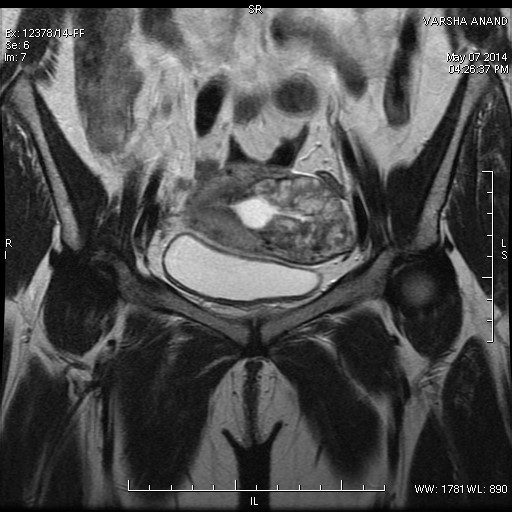
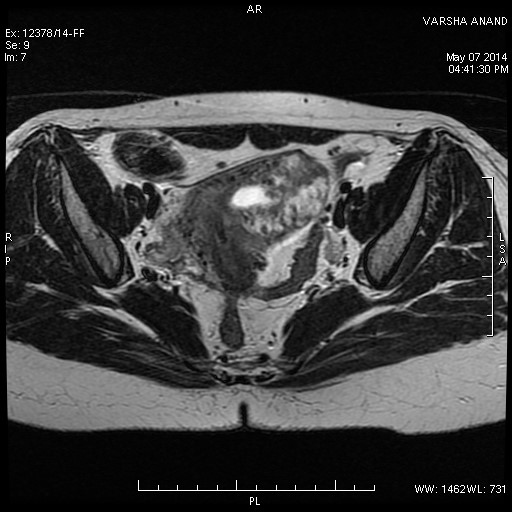
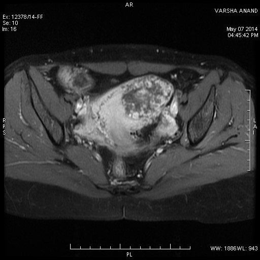
A young female had a history of recent D&C for missed abortion. She now presented with bleeding per vaginum and persistently elevated beta HCG levels. MRI Pelvis with contrast was performed.


Findings
Heterogeneously enhancing mass lesion is seen in the left uterine cornu involving the entire wall thickness. This represents a Gestational Trophoblastic Neoplasm in the given setting.
Discussion
Gestational trophoblastic neoplasms (GTN) are extremely rare pregnancy related tumors. These arise from the trophoblastic cells which normally form the placenta. The various forms of GTN include hydatiform mole, invasive mole and choriocarcinoma with increasing invasive and malignant potential. These tumors are sensitive to chemotherapy and generally have a good prognosis. They produce HCG hormone whose levels can be used to monitor tumor progression and response to therapy.
USG and MRI demonstrate the tumor as a heterogenous vascular mass in the uterus. MRI can clearly delineate the extent of uterine wall invasion and helps in local staging. Distant metastases are generally to the lung and brain and can be evaluated with CT Chest and MRI Brain respectively.
References :
- Nonmetastatic gestational trophoblastic neoplasia. Role of ultrasonography and magnetic resonance imaging (PMID:9475144). Kohorn EI, McCarthy SM, Taylor KJ. The Journal of Reproductive Medicine 1998, 43(1):14-20.
- Radiology of gestational trophoblastic neoplasia. S.D. Allena, A.K. Lima, M.J. Secklb, D.M. Blunta, A.W. Mitchella. Clinical Radiology Volume 61, Issue 4, April 2006, Pages 301–313.
