What is a Mammogram?
A mammogram is an X-ray examination of the breasts. A mammogram can help to diagnose any changes or symptoms in the breasts including lumps, tenderness or pain, nipple discharge or any skin changes.
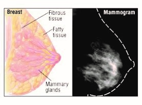
How it is done?
Firstly, we ask you to complete a questionnaire to obtain relevant information such as any family history of breast cancer, previous mammograms or breast surgery. We will provide you with gown. Your breasts will be placed, one at a time, between two special plates and compressed (pressed down) firmly for a few seconds whilst the x-ray are taken. Two views of each breast are performed as a minimum.
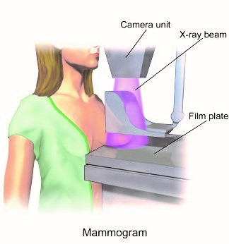
Is it painful?
The compression may be uncomfortable, or perhaps painful, but it only lasts a few seconds. Without compression, the x-rays would be blurry, which makes it difficult to see any abnormality. Compression also reduces the amount of radiation required for the mammogram. Ideally it is best to have a mammogram on week after the start of your menstrual cycle, as your breasts will not be as tender at this time.
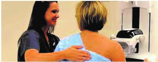

How long does it take?
Standard mammography takes approximately 15 minutes, as the images are reviewed by our Radiologists (a specialist Doctor with breast image training). Sometimes extra views are performed which take longer. If you have breast implants, the mammography will take longer (40 minutes), as extra images are needed in order to see the breast tissue clearly. An ultrasound is generally required after the mammogram which takes a further 30 minutes.
How do I Prepare?
Please don’t wear perfume, lotion or talcum powder on the day of your appointment, as these substances may show up as shadows on your mammogram. Bring your deodorant with you, so you put it on the end of the examination.
Wear a two piece of outfit, for comfort so you only need to undress from the waist up. Bring any previous mammogram films with you, so they can be compared with your mammogram.
If you have breast implants, please let us know when you are booking your appointment, as a longer appointment time will be scheduled.
If you are pregnant or breast feeding please let the Radiographer know before commencement of examination.
Breast Ultrasound:
The role of breast sonography is in the differentiation of solid & cystic lesions. Ultrasound compliments both clinical examination and mammography. It plays an important role in triple assessment of symptoms lesions in dense breast. It is the examination of choice in young woman and valuable in assessment of mammogram dense breasts.
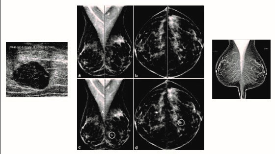
A mammogram is an x-ray of the breasts using low-dose radiation. The key role of mammography is identifying breast cancer early in its development when it is very small. This is often a year or two before it is large enough to be felt by you or your healthcare provider as a lump. A screening mammogram is used to help find breast cancer early in women who have no symptoms. With a screening mammogram, a radiologist is not only looking for calcifications, cysts and fibroadenomas (solid lumps of normal breast cells). A diagnostic mammogram may be done as a problem-solving examination in patients who have abnormal physical findings or an abnormal screening mammogram. Diagnostic mammograms may also be used for patients with breast implants.
Mammography detects about 2 to 3 times as many early breast cancers as a physical examination and is considered the “gold standard” in breast cancer detection. It is the best method to screen for the presence of a small undetectable lump or a group of micro-calcifications, which may be the only sign of breast cancer. While mammography is the best screening exam available today, about 1 in 10 cancers will not be identified until they can felt as lumps. That is why breast self-examination and regular exams by your healthcare provider remain integral components of breast cancer detection.
CANCER FACTS
- Breast cancer is the most common cancer in women, aside from skin cancer.
- Breast cancer is the second leading cause of cancer death, after lung cancer in women.
- One in every eight women in the United States will develop breast cancer in her lifetime.
- This year, more than 2,00,000 new cases of invasive breast cancer will be diagnosed among women in our country.
Currently, two-thirds of breast cancers are diagnosed at localized stage, for which the five-year survival rate is 97%. This high rate of early detection can be attributed to utilization of mammography screening. Better understanding of breast cancer symptoms within our population has also helped to increase detection.
