When diagnosing injuries or internal conditions, doctors often rely on imaging tools. Two of the most common are the MRI scan and the X-ray. While both provide valuable insights, they differ in technology, detail, and use. This guide explains what an MRI scan shows that an X-ray cannot—and when you might need one over the other.
What Is an MRI Scan?
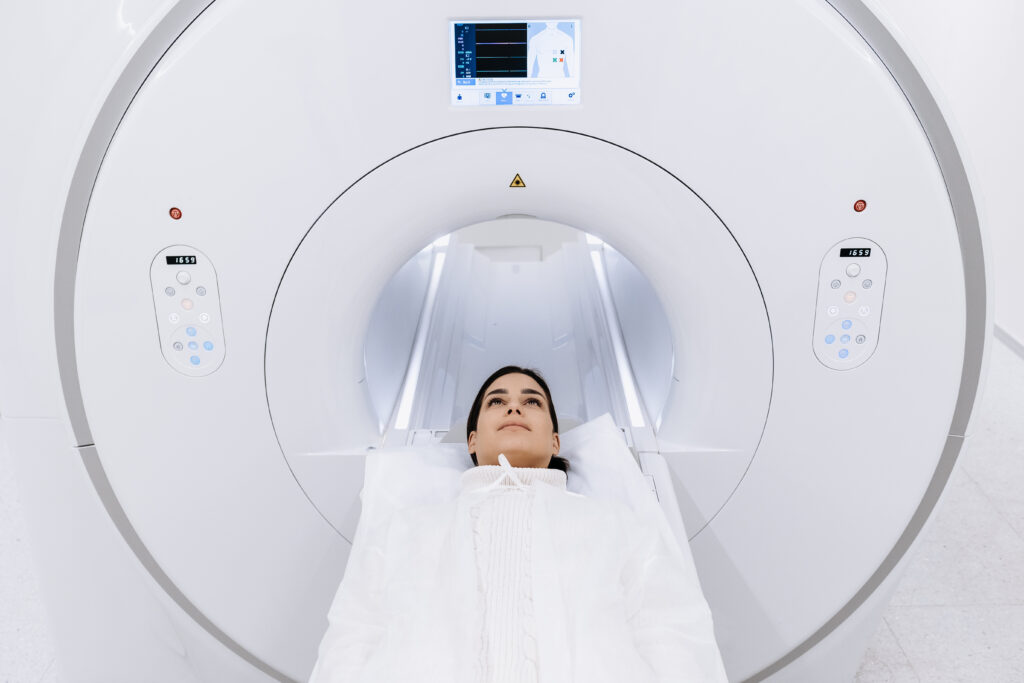
An MRI scan (Magnetic Resonance Imaging) uses strong magnets and radio waves to produce detailed images of your internal body structures. It’s especially effective for viewing soft tissues like the brain, muscles, and organs. One of the key MRI scan benefits is that it does not use harmful radiation.
What Is an X-Ray?
An X-ray uses ionizing radiation to capture images of the inside of the body. It is fast, affordable, and ideal for viewing bones. However, it’s not as effective for diagnosing soft tissue conditions.
MRI Scan vs. X-Ray: Key Differences
Feature
MRI Scan
X-Ray
Technology
Magnetic fields & radio waves
Ionizing radiation
Radiation
No
Yes
Image Detail
High (best for soft tissues)
Moderate (best for bones)
Use Case
Brain, joints, organs, spinal cord
Bones, fractures, lungs
Time Required
15–60 minutes
Few seconds
Comfort Level
Requires staying still
Quick and simple
Cost
Higher
Lower
What an MRI Scan Shows That an X-Ray Does Not
1. Soft Tissue Injuries
MRI scans clearly show ligaments, tendons, cartilage, and muscles—details an X-ray cannot capture.
2. Brain & Spinal Cord
For detecting brain tumors, stroke, multiple sclerosis, or disc issues in the spine, an MRI scan is far superior.
3. Joint & Cartilage Damage
If you’re suffering from knee pain or shoulder injuries, an MRI scan can detect meniscus or rotator cuff tears, unlike an X-ray.
4. Internal Organs
MRI scans can detect tumors or inflammation in the liver, kidneys, uterus, and other soft tissues that an X-ray would miss.
5. Blood Flow and Vessels
MRI angiography maps blood vessels without dye or radiation, making it a powerful diagnostic tool not available with a standard X-ray.
MRI Scan vs. X-Ray: Pricing Comparison
At Ace Imaging Centre
Service Type
Scan Type
Price (INR)
MRI Scan
Brain
₹8,000
Spine
₹7,500
Knee
₹6,000
Shoulder
₹6,500
Full Body
₹15,000
X-Ray
Chest
₹500
Limb
₹300
Spine
₹700
Abdomen
₹600
Note: Prices are indicative and subject to change.
MRI Scan Benefits
No Radiation Exposure – Safe for repeat use.
Detailed Imaging – Especially useful for soft tissue, nerves, and organs.
When Should You Choose an MRI Scan Over an X-Ray?
Soft tissue injuries (e.g., ligaments, tendons)
Brain or spinal issues
Chronic joint pain
Organ abnormalities
Follow-up imaging for unclear X-ray results
Choose an X-ray when:
Bone fractures or dislocations are suspected
You need quick and low-cost imaging
Lung issues like pneumonia are suspected
FAQs
1. What can an MRI scan detect that an X-ray can’t?
Soft tissue damage, brain disorders, and internal organ issues.
2. Is an MRI scan safe?
Yes, it uses no radiation and is non-invasive.
3. When is an X-ray better?
For viewing bones, fractures, or chest conditions quickly.
4. Which is more detailed—MRI scan or X-ray?
MRI scan offers more detailed images of soft tissues.
5. Is an MRI scan more expensive than an X-ray?
Yes. MRI scans are costlier due to advanced technology.
Conclusion
While X-rays are fast and effective for bones, MRI scans provide a deeper view of soft tissues, organs, and the brain. Knowing what each scan offers helps you and your doctor choose the best option for your diagnosis. If you need high-quality imaging services, Ace Imaging Centre offers expert support, advanced equipment, and competitive pricing.
When your doctor suggests a scan to better understand what’s happening inside your body, you’ll likely encounter two major options: CT (Computed Tomography) scans and MRI (Magnetic Resonance Imaging) scans. Both are powerful diagnostic tools used to produce detailed internal images. But they differ significantly in terms of how they work, what they are best suited for, their safety profiles, and cost.
In this comprehensive guide, we’ll explore the difference between CT scan and MRI, including their uses, advantages, limitations, and the affordable scan pricing offered at Ace Imaging Centre. If you’re wondering about CT scan vs MRI for your condition, this guide will help you understand what suits your needs best.
Understanding the Basics
What Is a CT Scan?
A CT scan uses X-rays to capture detailed cross-sectional images of the body. Unlike a regular X-ray, a CT scan rotates around your body to take multiple images from different angles. These images are then processed by a computer to create 3D representations of bones, organs, and tissues.
CT scans are widely used in hospitals and clinics, particularly in emergency settings, because they are fast and efficient.
Key Features of CT Scans:
Uses ionizing radiation (X-rays)
Fast (usually 5–10 minutes)
Good for visualizing bones, chest, lungs, and abdominal structures
Often used in trauma or emergency care
What Is an MRI Scan?
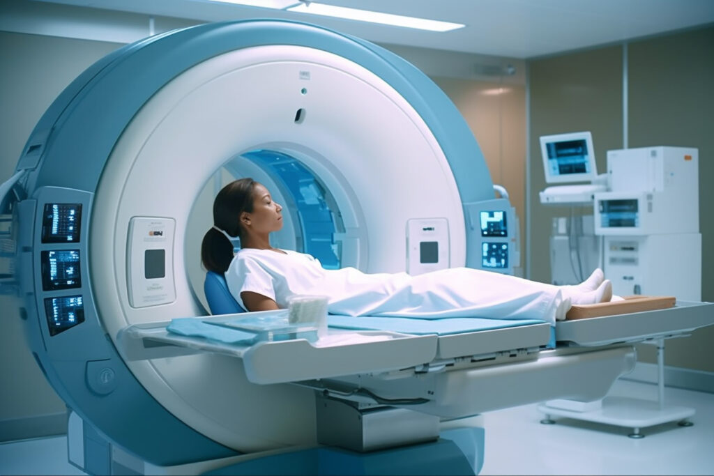
An MRI scan uses magnetic fields and radiofrequency pulses to produce detailed images of internal organs and tissues. Instead of X-rays, MRI machines use a strong magnet and radio waves that interact with hydrogen atoms in the body to generate images.
MRIs take longer to perform but provide superior contrast when examining soft tissues.
Key Features of MRI Scans:
No ionizing radiation
Takes 30–60 minutes
Provides highly detailed soft tissue images
Ideal for brain, spinal cord, joints, and muscles
How Do CT and MRI Scans Work?
Understanding the core technology behind these scans helps clarify their ideal applications.
Technology Behind CT Scans
CT scans emit X-rays through the body, capturing multiple slices or cross-sections that are assembled into a 3D image. The X-ray beams are absorbed differently by various tissues (bone, fat, fluid), enabling the scan to highlight abnormalities.
This is especially effective for:
Fractures
Internal bleeding
Tumors
Lung diseases
Abdominal injuries
Technology Behind MRI Scans
MRI scans rely on magnetic fields and radio waves to detect the natural energy emitted by hydrogen atoms in the body. When exposed to magnetic fields, these atoms align and produce signals that are captured and converted into images by the MRI machine.
MRIs are preferred when doctors need a more detailed look at:
Brain and spinal cord
Joints and ligaments
Tumors in soft tissue
Muscles and internal organs
CT Scan vs MRI: Comparative Overview
To make the difference between CT scan and MRI clearer, here’s a side-by-side comparison:
Aspect
CT Scan
MRI Scan
Technology
X-rays
Magnetic fields + radio waves
Radiation
Yes (low dose)
No
Time Required
5–10 minutes
30–60 minutes
Best for
Bones, lungs, trauma, internal bleeding
Brain, spine, joints, soft tissues
Image Detail
Moderate (excellent for bone & lungs)
High (excellent for tissues & nerves)
Cost
Lower
Higher
Noise Level
Quiet
Loud (ear protection provided)
Safety with Implants
Usually safe
Not suitable for patients with metal implants
Contrast Dye
Iodine-based (may cause allergies)
Gadolinium-based (safer, but check kidney health)
If you’re comparing a CT scan vs MRI, this table offers a helpful reference based on technology, safety, application, and cost.
When to Choose a CT Scan
CT scans are ideal in situations where speed and accuracy are critical, particularly in emergency or trauma care. Doctors prefer CT scans when they need quick answers to:
Detect internal injuries or bleeding
Evaluate chest pain or stroke symptoms
Diagnose complex bone fractures
Assess cancer spread
Guide biopsies or surgeries
Advantages of CT Scans:
Fast and widely available
Excellent for bone and chest imaging
Can capture moving structures (e.g., lungs)
Limitations:
Exposure to radiation
Less detail in soft tissues compared to MRI
When to Choose an MRI Scan
MRI scans provide excellent detail for soft tissues, nerves, and the brain. They are often used in planned, non-emergency scenarios when physicians need detailed information to diagnose or monitor complex conditions.
Ideal for Diagnosing:
Brain tumors, strokes, or multiple sclerosis
Spinal cord injuries and disc herniation
Ligament tears and sports injuries
Soft tissue tumors or inflammation
Pelvic or abdominal organ conditions
Advantages of MRI Scans:
No radiation exposure
Highly detailed imaging of soft tissues
Safe for repeated use
Limitations:
Longer scan time
Claustrophobia or discomfort due to noise
Incompatibility with some implants (e.g., pacemakers)
MRI vs CT Scan Cost in India
One major consideration for patients is cost. While MRI scans are more expensive due to the complex technology and longer time, both scans remain essential tools in medical diagnostics.
Affordable Imaging at Ace Imaging Centre
Scan Type
Price
MRI Scan
₹3,999
CT Scan (Plain)
₹1,500
CT Chest (Plain)
₹2,500
CT Abdomen (Plain)
₹3,500 (Contrast extra)
Ace Imaging Centre offers competitive rates without compromising on quality. We use state-of-the-art equipment and have experienced radiologists on staff to ensure reliable, fast results. Whether it’s a CT scan vs MRI decision or a general diagnostic need, you’ll find high-quality services at affordable prices here.
What to Expect During the Scans
During a CT Scan:
You’ll lie on a table that moves through a donut-shaped scanner.
The process is painless and quick.
You may be asked to hold your breath for a few seconds.
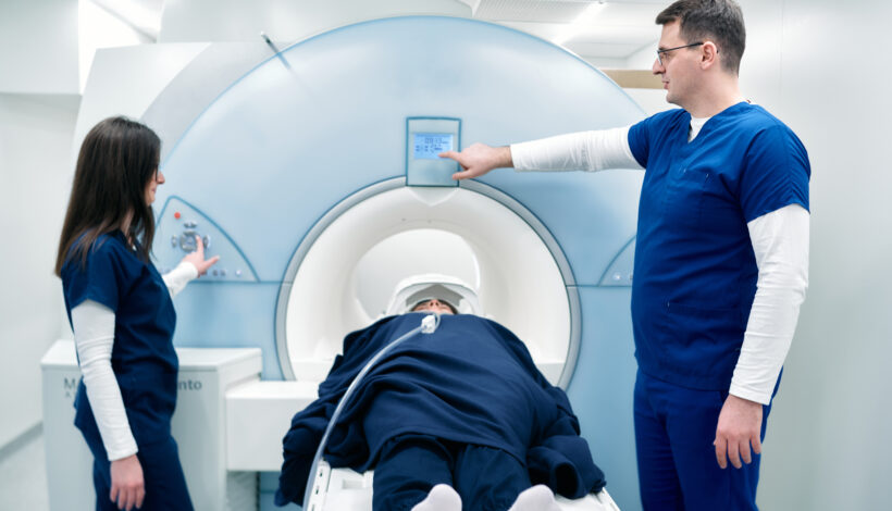
During an MRI Scan:
You’ll lie on a narrow table that slides into a tube-like machine.
The scan is noisier, and you’ll be given earplugs or headphones.
You must remain still for the best image quality.
The experience is painless but may feel long for some.
Safety and Precautions
CT Scan Safety:
Generally safe for most people.
Not recommended for pregnant women due to radiation.
Rare allergic reactions to contrast dye.
MRI Safety:
No radiation involved.
Safe for most patients unless they have metal implants, pacemakers, or certain tattoos.
Gadolinium contrast is usually safe but not recommended for patients with severe kidney disease.
Always inform your doctor and technician about existing health conditions, implants, allergies, or pregnancy before undergoing any imaging procedure.
FAQs
1. Which scan is more detailed—CT or MRI?
MRI is more detailed for soft tissues, nerves, and the brain. CT is better for bones and chest imaging.
2. Is an MRI safer than a CT scan?
Yes. MRI is safer for repeated use as it does not involve radiation. CT scans involve a small dose of radiation.
3. How long does each scan take?
CT scans take 5–10 minutes, while MRI scans usually take 30–60 minutes depending on the body part.
4. Can I get a scan with a metal implant?
CT scans are generally safe with implants. MRI may not be suitable if you have pacemakers, aneurysm clips, or certain metallic implants.
5. Do I need to fast before my scan?
Not always. Fasting is required only for certain contrast-enhanced scans. Your doctor will guide you based on the scan type.
Why Choose Ace Imaging Centre?
At Ace Imaging Centre, we are dedicated to providing accurate diagnostics, patient comfort, and affordable care. Our facility is equipped with the latest MRI and CT scan machines, and our team is committed to fast, precise reporting.
What We Offer:
Expert radiologists
Same-day reporting
Affordable scan packages
Patient-friendly environment
Online appointment scheduling
Whether you need a quick CT scan or a detailed MRI scan, our team is here to help you every step of the way.
Conclusion
Understanding the difference between CT scan and MRI empowers you to make informed decisions about your health. Both imaging technologies are essential in modern diagnostics, and the best choice depends on your condition and what your doctor needs to evaluate.
At Ace Imaging Centre, we provide high-quality, affordable diagnostics for all patients. If you’re uncertain about CT scan vs MRI, our experts are ready to assist.
Book your scan today or call us for more information on how we can support your health journey.
When it comes to diagnostic imaging, two of the most commonly recommended tests are MRI (Magnetic Resonance Imaging) and CT (Computed Tomography) scans. While both are used to look inside the body, they differ in technology, use cases, and — most importantly for many patients — cost.
If you’re trying to decide between these two scans or you’ve been advised by a healthcare provider to get one, understanding the MRI vs CT scan cost differences can help you make an informed choice. This guide breaks it all down — from how the scans work to how much they cost at Ace Imaging Centre.
Table of Contents
“MRI vs PET Scan: A visual guide to understand key differences in medical imaging”
What is an MRI Scan?
An MRI (Magnetic Resonance Imaging) scan uses powerful magnetic fields and radio waves to create detailed images of the organs and tissues in your body. It is a non-invasive and radiation-free procedure ideal for:
Internal organ scans (like the liver, kidneys, or uterus)
Key Features of MRI:
No radiation exposure
Longer scan times (30–60 minutes)
High-resolution images of soft tissues
Not suitable for patients with metal implants or pacemakers
What is a CT Scan?
A CT (Computed Tomography) scan uses X-ray technology to capture multiple images from different angles, which are then compiled to form cross-sectional views of the body. It’s often the go-to imaging tool in emergency settings.
CT Scans Are Commonly Used For:
Diagnosing bone fractures
Detecting internal injuries and bleeding
Evaluating lung and chest conditions
Identifying tumors or masses
Planning surgeries or treatments
Key Features of CT:
Quick scan times (5–10 minutes)
Involves low-dose radiation
Excellent for imaging bones and chest
May involve contrast dyes for better clarity
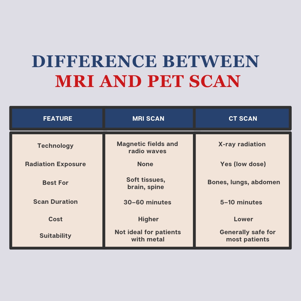
MRI vs CT Scan: Key Differences
Feature
MRI Scan
CT Scan
Technology
Magnetic fields and radio waves
X-ray radiation
Radiation Exposure
None
Yes (low dose)
Best For
Soft tissues, brain, spine
Bones, lungs, abdomen
Scan Duration
30–60 minutes
5–10 minutes
Noise Level
Loud (requires ear protection)
Minimal noise
Suitability
Not ideal for patients with metal
Generally safe for most patients
Cost
Higher
Lower
The MRI vs CT scan cost is primarily influenced by the complexity of the imaging process, technology used, and the duration of the scan. MRI is typically more expensive due to the advanced technology and longer scan times.
MRI vs CT Scan Cost at Ace Imaging Centre
At Ace Imaging Centre, we believe in transparency and affordability. Below is a breakdown of our current imaging prices:
💰 Imaging Pricing Table
Scan Type
Price (INR)
MRI Scan
₹3,999
CT Scan (Plain)
₹1,500
CT Chest (Plain)
₹2,500
CT Abdomen (Plain)
₹3,500 (Contrast extra)
These prices reflect high-quality diagnostic imaging with the latest technology, performed by expert radiologists. Compared to market averages, Ace Imaging Centre offers competitive rates without compromising on accuracy or patient care.
Which Scan Should You Choose?
If your doctor has not specified which scan is required, understanding the purpose behind each can help you discuss your options better.
There’s a need to evaluate joint damage or spinal discs
Choose CT if:
You need a fast diagnosis, such as in trauma cases
You’re being evaluated for chest or abdominal issues
Bone fractures or internal bleeding are suspected
Still unsure? Our radiology team at Ace Imaging Centre is here to guide you on what’s best for your health condition and budget.
MRI vs CT Scan Cost: Why the Difference?
The cost of MRI vs CT scan varies due to several key factors:
Equipment Costs: MRI machines are more expensive to operate and maintain.
Scan Duration: MRI takes longer, requiring more technician time and scheduling logistics.
Image Detail: MRI provides greater clarity for soft tissue structures.
Radiation-Free: MRI uses advanced magnetic technology, which increases cost but reduces long-term health risks.
When weighing MRI vs CT scan cost, always consider the type of information needed for accurate diagnosis. A lower-cost scan might not provide the detail your physician needs, which could lead to repeated or follow-up imaging.
Frequently Asked Questions
1. What is the cost of MRI scan?
At Ace Imaging Centre, the cost of an MRI scan is ₹3,999. This is an all-inclusive rate for a standard MRI scan, providing high-quality imaging with no hidden fees. Our advanced MRI machines ensure optimal clarity and comfort during the procedure.
2. Is an MRI more expensive than a CT scan?
Yes, MRI scans are typically more expensive than CT scans. At Ace Imaging Centre:
MRI scan costs ₹3,999
CT scans start from ₹1,500
The price difference is due to the advanced technology, longer scan time, and the type of imaging data produced.
3. Can I choose which scan to take based on cost alone?
While cost is an important consideration, the choice between MRI and CT should be made based on medical necessity. If you’re unsure, consult your referring doctor or speak to our radiology team for personalized guidance.
4. Do prices differ for contrast scans?
Yes. For example, CT Abdomen (Contrast) costs more than the plain scan. Contrast-enhanced scans require additional materials and monitoring, which increase the total cost. Please contact Ace Imaging Centre for a detailed quote if your doctor has recommended a contrast study.
5. Is prior preparation needed for these scans?
MRI: May require fasting, especially for abdominal scans. You’ll need to remove metal objects and inform the technician about implants.
CT: Often requires no special preparation unless contrast is involved. Hydration before and after the scan is encouraged if contrast is used.
Conclusion
Understanding the MRI vs CT scan cost is essential for patients looking to make informed, budget-conscious healthcare decisions. While MRI scans offer greater detail for soft tissues, they do come at a higher price. CT scans, on the other hand, are quicker and more affordable, making them ideal for many urgent diagnostic needs.
At Ace Imaging Centre, we offer both MRI and CT services at competitive prices, combining accuracy, safety, and compassionate care. Whether you’re booking your first scan or seeking a second opinion, our expert team is ready to assist.
In the evolving world of medical imaging, accuracy and early detection are critical—especially when it comes to diagnosing complex conditions like neuroendocrine tumors (NETs). Among the most advanced tools available today is the DOTA scan, a specialized PET scan that helps doctors see beyond what traditional imaging methods can reveal.
But what exactly is a DOTA scan, and why are so many specialists recommending it for diagnosis and follow-up care? In this blog, we’ll dive into what makes a DOTA scan so beneficial, when it’s used, and how it can change the course of treatment planning for the better.
What Is a DOTA Scan?
A DOTA scan commonly called a Ga-68/ 18-F DOTA PET scan is a highly sensitive imaging technique used to detect tumors that express somatostatin receptors, primarily neuroendocrine tumors. The scan involves using a radiopharmaceutical agent, often labeled with Ga-68/ 18-F, which binds specifically to these receptors and highlights abnormal tissue during the scan.
Unlike regular PET or CT scans, a DOTA scan provides clearer, more precise information about tumor location, size, and activity, making it a critical diagnostic tool.
Top Benefits of DOTA Scan
1. Highly Accurate Tumor Detection
One of the main reasons doctors recommend a DOTA scan is because of its unmatched accuracy. It can detect small tumors that might be missed by CT or MRI. This is particularly crucial for neuroendocrine tumors that are often slow-growing and hard to locate.
2. Early and Precise Diagnosis
Early detection is key to effective treatment. A DOTA scan can identify disease at an earlier stage than many other imaging methods. This early insight gives oncologists a head start in developing a targeted treatment plan.
3. Better Staging of Cancer
Knowing how far a tumor has spread is essential. The DOTA scan offers detailed images that help doctors accurately stage cancer, which directly impacts treatment decisions such as surgery, chemotherapy, or PRRT (Peptide Receptor Radionuclide Therapy).
4. Guides Personalized Treatment Plans
With its detailed imaging, the DOTA scan plays a crucial role in mapping out personalized treatment strategies. Doctors can see which areas are active and tailor therapies accordingly, especially for patients undergoing PRRT or monitoring tumor progression.
5. Effective for Follow-Up Monitoring
A DOTA scan isn’t just useful during diagnosis—it’s also vital during follow-up. Patients with known neuroendocrine tumors often undergo periodic scans to ensure the tumor hasn’t returned or progressed. The high sensitivity of DOTA imaging helps catch even minor changes.
6. Low Radiation Exposure Compared to Traditional Imaging
Despite its powerful capabilities, the DOTA scan uses relatively low radiation. This makes it safer for repeated use, especially in patients who require regular follow-ups or monitoring over several years.
7. Quick and Comfortable Procedure
Most DOTA scans are completed within a few hours, and the procedure is non-invasive. Patients usually receive a small injection of the radiotracer and rest before being scanned. The actual scan takes about 20–30 minutes and is generally painless.
When Is a DOTA Scan Recommended?
Doctors typically recommend a DOTA scan in the following scenarios:
When a patient presents symptoms suggestive of neuroendocrine tumors (NETs)
For accurate cancer staging and restaging
To evaluate eligibility for PRRT therapy
During routine follow-ups to detect recurrence or metastasis
In patients with a previous inconclusive scan result from CT or MRI
Because of its specificity, the DOTA scan is often preferred over traditional octreotide scans and sometimes even over PSMA PET scans for certain cancers.
Why Choose Ace Imaging Center for Your DOTA Scan?
When it comes to high-precision imaging, Ace Imaging Center stands out as a leading destination for DOTA scans in Mumbai. Equipped with advanced PET-CT technology and a team of expert radiologists, Ace Imaging ensures quick, accurate, and patient-friendly diagnostic services. The center follows strict safety protocols and provides detailed reports that assist your oncologist in making timely decisions. Whether it’s your first DOTA scan or a follow-up, Ace Imaging is committed to delivering top-notch care, clarity, and convenience at every step.
Final Thoughts
In summary, the DOTA scan has transformed the way doctors approach cancer diagnosis and treatment—especially for patients with neuroendocrine tumors. Its high precision, safety profile, and utility in both diagnosis and follow-up make it a trusted imaging choice among oncologists worldwide.