Medical imaging plays a vital role in diagnosing, monitoring, and managing a wide range of conditions—from cancer and neurological disorders to internal injuries and infections. Two of the most widely used diagnostic tools are the PET scan and CT scan. Though they are often mentioned together, these scans serve different purposes and work in unique ways.
In this guide, we’ll walk you through the basics of each scan, what to expect before, during, and after your appointment, and help you better understand which one might be recommended for you.
Understanding the Scans
| Feature | CT Scan | PET Scan |
| Full Name | Computed Tomography | Positron Emission Tomography |
| Technology Used | X-rays to create cross-sectional images | Radioactive tracers to observe metabolic activity |
| Purpose | Shows structural detail | Shows functional processes |
| Common Uses | Bone injuries, tumors, blood clots, organ structure | Cancer detection, heart conditions, brain disorders |
| Scan Duration | 5–10 minutes | 30–60 minutes (including tracer time) |
| Radiation Involved | Yes | Yes (with tracer) |
| Contrast Material | Often used | May not be needed |
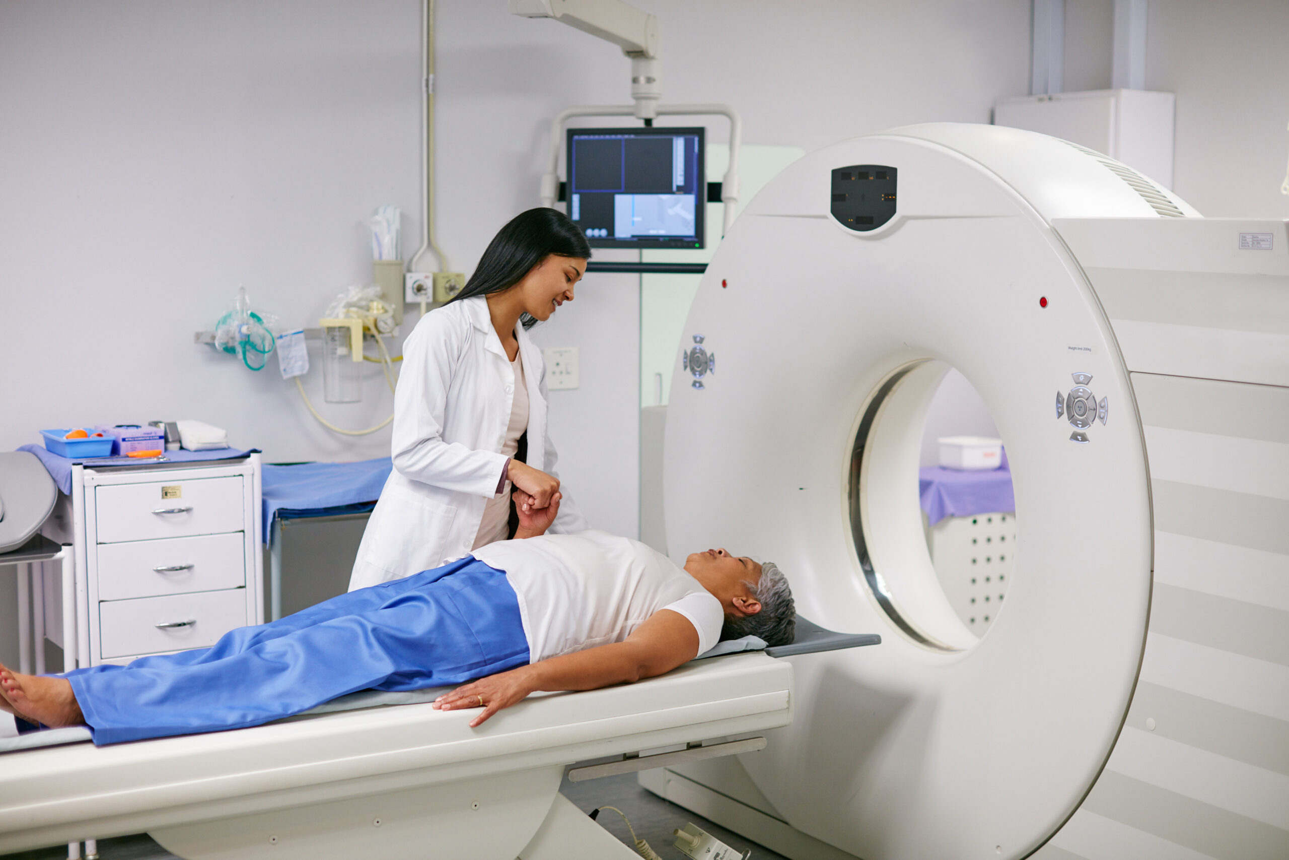
What Is a CT Scan?
A CT scan is a diagnostic tool that combines X-rays and computer technology to produce detailed images of bones, organs, and soft tissues. It is commonly used for detecting:
- Internal bleeding or injuries
- Lung and liver conditions
- Bone fractures
- Tumor detection
- Monitoring treatment response in cancer
At Ace Imaging Center, our advanced CT machines produce high-resolution images quickly and with minimal discomfort. The scan is typically short and non-invasive, making it a preferred choice for many urgent and routine cases.
What Is a PET Scan?
A PET scan uses a small amount of radioactive material called a tracer, which is injected into the bloodstream. The tracer travels through your body and highlights areas with increased metabolic activity—commonly found in cancer cells or certain brain and heart disorders.
Conditions PET scans can help diagnose and monitor include:
- Cancer (diagnosis, staging, recurrence)
- Alzheimer’s and other neurological conditions
- Fever of unknown origin – to identify hidden focus of infection in entire body.
- Diagnosing Inflammatory conditions like sarcoidosis or vasculitis
- Epilepsy and seizure mapping
The scan itself is painless. However, you’ll need to rest for a while after the tracer is injected, as your body absorbs it before the actual imaging takes place.
What to Expect: Before, During, and After the Scan
Here’s a clear, side-by-side comparison of what you can expect at each stage of the imaging process:
| Stage | CT Scan | PET Scan |
Before | – May be asked to fast for 2–4 hours if contrast is used. | – Fasting for 4–6 hours may be required. |
| – Inform if you have allergies, kidney issues, or are pregnant. | – Avoid strenuous activity the day before. Inform if diabetic or pregnant. | |
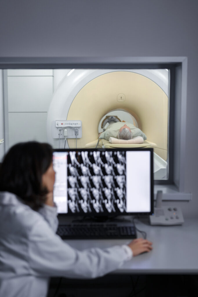
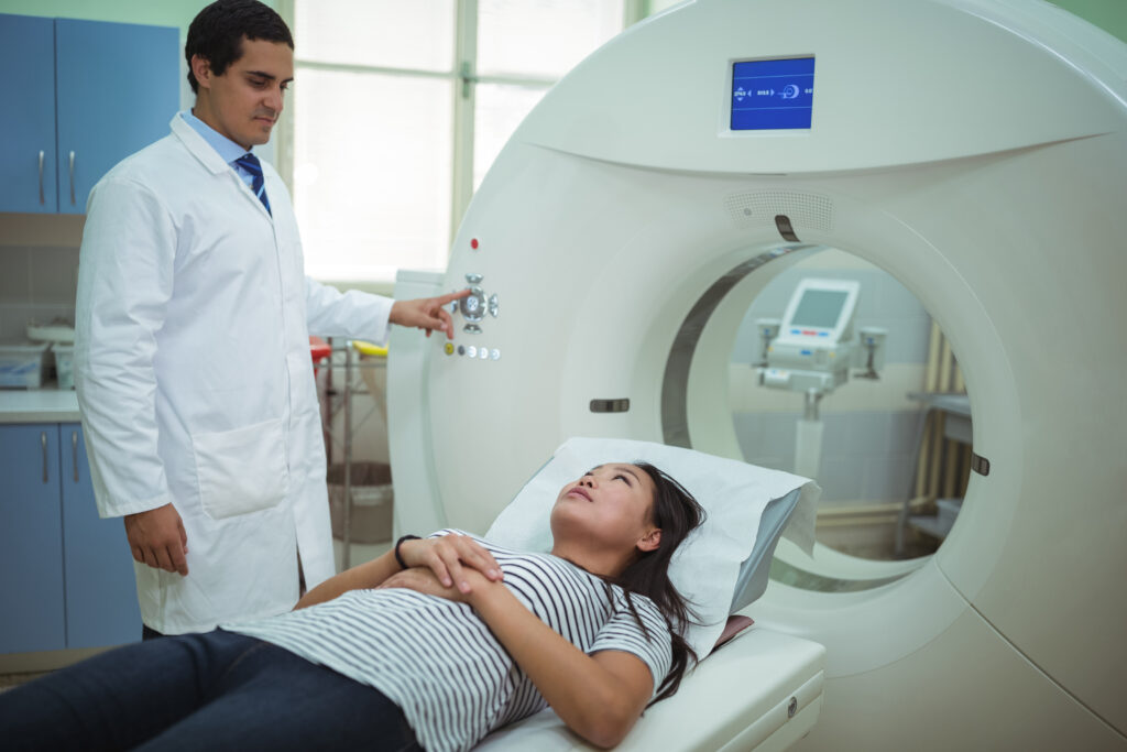
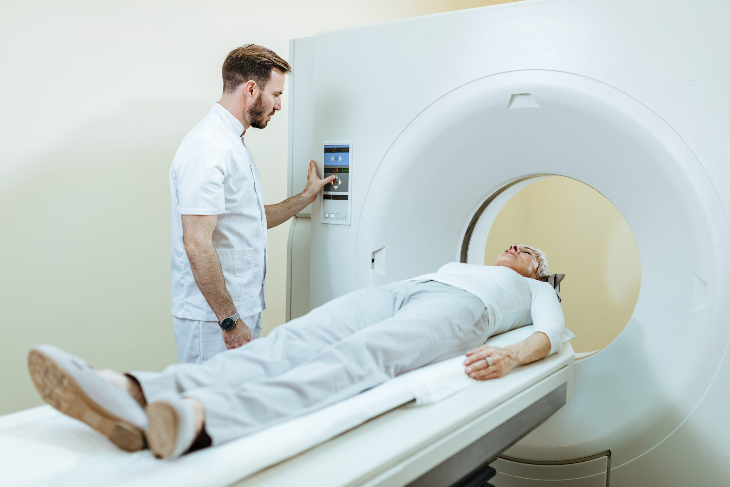
During | – You’ll lie on a table that slides through a doughnut-shaped scanner. | – A small injection of tracer is given first. You’ll rest quietly for 30–60 minutes. |
| – Scan time is typically 5–10 minutes. | – You’ll then lie still on the scan table for 20–30 minutes while images are taken. | |
After | – You can usually return to normal activities right away. | – Drink plenty of water to help flush out the tracer. |
| – If contrast was used, drink fluids to help eliminate it from your body. | – Avoid close contact with infants or pregnant women for several hours post-scan. |
Is It Safe?
Both scans involve exposure to a small amount of radiation. However, the benefits of accurate diagnosis often far outweigh the risks. These imaging tools are approved by medical authorities and used globally with great success.
At Ace Imaging Center, we follow strict safety protocols and use low-dose techniques wherever possible to ensure your well-being. Our team will explain the process and answer all your concerns before your scan.
Patient Comfort at Ace Imaging Center
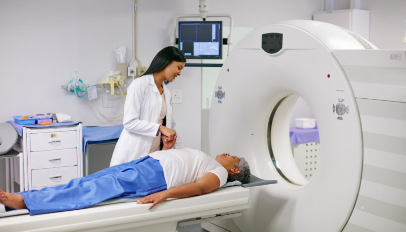
At Ace Imaging Center, we prioritize patient care at every step. From the moment you arrive, our team works to make you feel comfortable and informed. We maintain a clean, peaceful environment and use state-of-the-art machines that provide faster, more accurate results with minimal discomfort.
Whether it’s your first scan or a follow-up, we understand that diagnostic imaging can feel overwhelming. Our technicians and radiologists take the time to walk you through the process and ensure that you’re confident and at ease throughout your visit.
Conclusion
When it comes to choosing between a PET scan vs CT scan, the decision depends on what your doctor needs to diagnose or monitor. A CT scan provides detailed anatomical images perfect for detecting physical injuries, tumors, or organ conditions. A PET scan, on the other hand, focuses on metabolic activity, helping detect diseases at a cellular level, even before structural changes appear.
At Ace Imaging Center, we offer both PET and CT scanning under one roof, along with expert guidance to help you and your doctor make informed choices. With cutting-edge imaging equipment, trained professionals, and a patient-first approach, we’re committed to providing you with accurate, timely, and compassionate diagnostic care.